NANOSPACER contributes to cancer research
NANOSPACER is published in the International Journal of Nanomedicine, for contributions to cancer research in collaboration with University of Bergen.
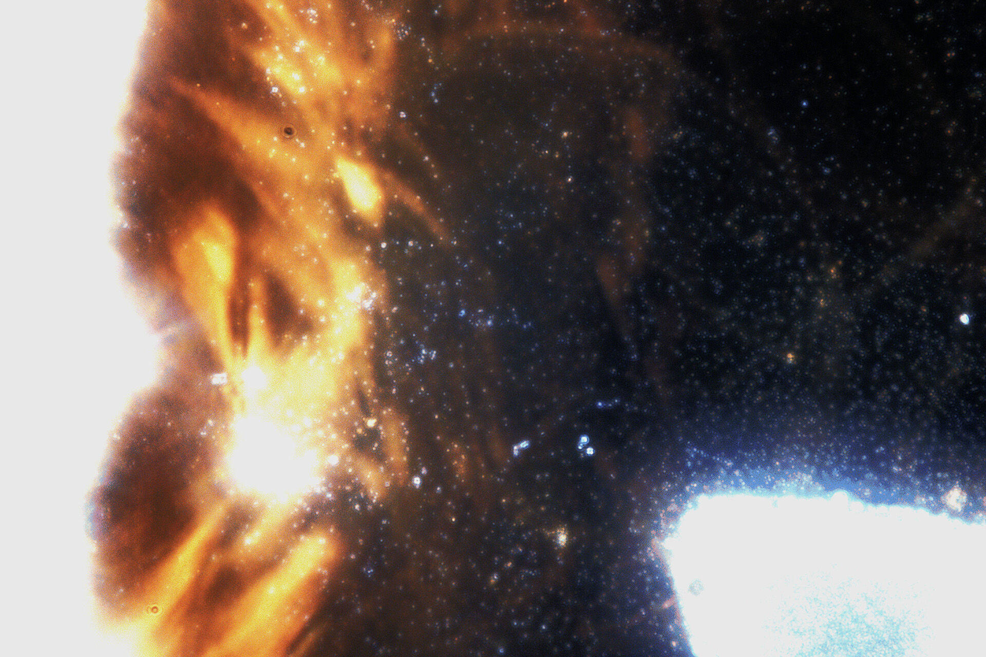
Magnetic iron-oxide nanoparticles (orange/red) and plastic nanoparticles (blue) imaged in NANOSPACER. (Illustrative purposes only. Image colour is real and can be seen with the bare eye.) Image by Oliver Vanderpoorten.
Researchers from Bergen Haukeland University Hospital, Qilu Hospital of Shandong University, and Shandong Key Laboratory of Brain Health and Function Remodeling have demonstrated a new method to label brain-metastasis-derived EVs using superparamagnetic iron oxide nanoparticles (SPIONs) which enables their visualization and tracking through magnetic resonance imaging (MRI) - even in vivo!
NANOSPACER was a key contribution to validate cargo-uptake (SPIONs) of extracellular vesicles and correlated with measurements taken with golden standard methods such as electron microscopy (EM) and dynamic light scattering (DLS).
“This is the first application of the technology and a big step for open-source NTA (Nanoparticle Tracking Analysis) and I hope the method finds more users in the field of biology, medicine and biotechnology,” says Oliver Vanderpoorten, researcher at UiT.
Read the article here
Journal of Nanomedicine:
Multimodal imaging of brain metastasis-derived extracellular vesicles | IJN
When cancer spreads to the brain, tiny particles called extracellular vesicles (EVs) play a key role. These particles, released in large numbers by tumor cells, carry signals that prepare the brain for cancer’s arrival by weakening natural defenses and altering the environment. Scientists are still learning how these brain metastasis EVs (BM-EVs) travel and interact with brain cells. Tracking them is difficult, especially with standard imaging tools like MRI, which can’t detect such small particles without help. That is where NANOSPACER comes in!
Published June 20, 2025 4:10 PM
- Last modified June 20, 2025 11:00 PM
