

Typically doctors use a technique called histological staining and microscopy to get a closer look at tissues and check for problems. However, these methods aren't always sensitive enough to spot subtle changes in the matrix. It's like trying to see the details of a fine painting with foggy glasses. The dyes used in traditional staining can make things even blurrier, and this makes it complicated to get an accurate assessment.
A superpower for microscopes
But fear not! We have devised a clever solution called MUSICAL, which is short for Multiple Signal Classification. It's like a superpower for microscopes, letting us see things that were invisible before. But that's not all! We've taken MUSICAL to the next level with a method called MUSI-tAF. With MUSI-tAF, we can zoom in on proteins like collagen in tissue samples. This allows detailed examination without the need for fancy equipment or years of training. This groundbreaking, label-free super-resolution method can reveal nanostructural details and density variations across a wide area of dense tissue matrix proteins. It's like having a microscope that can see way smaller!
Taking advantage of autofluorescence
MUSI-tAF works by using low magnification objectives and takes advantage of the natural glow emitted by proteins like collagen when exposed to light. By combining this autofluorescence with our powerful algorithm, we can now resolve even the tiniest structures within the matrix. This means we can uncover nanostructural details and variations in density between tissues undergoing fibrotic maladaptations which could catch problems before they become serious.
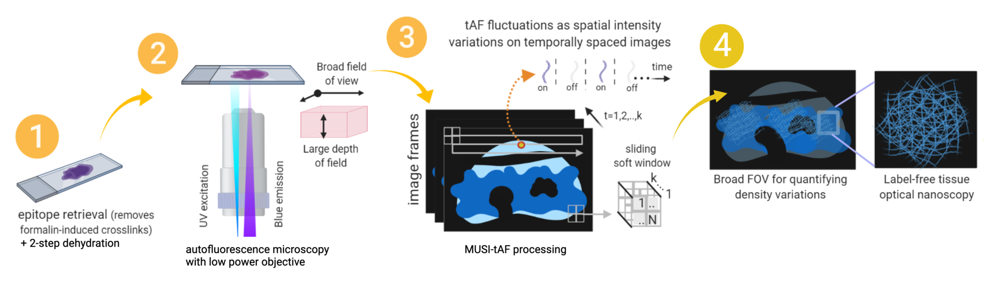
To validate this approach, we employed MUSI-tAF to identify clinically relevant features in various stages of pre-cancer, fibrotic diseases, and healing scars. And the results were impressive! MUSI-tAF was able to spot fine changes in the matrix that traditional methods might have missed, enhancing a doctor’s capability to diagnose and treat diseases early.
A valuable addition
Tissue matrix integrity and distribution plays a key role in numerous diseases. Incorporating MUSI-tAF into routine histopathological evaluations would be valuable addition to the arsenal of existing pathological practices for label-free molecular diagnosis.
So, thanks to MUSI-tAF, the future of diagnosing matrix-related diseases is looking more promising than ever. Making use of this tool, it’s easy to see how we could catch problems sooner and potentially improve patient outcomes.
To read more here is our published article:
Ghosh, B. et al. Molecular histopathology of matrix proteins through autofluorescence super-resolution microscopy. Sci Rep 14, 10524 (2024).