nanoRIP
nanoRIP
Combining optical and mathematical systems to create a better nanoscope.
Project overview
Project lead: Krishna Agarwal
Institution: University of Tromsø – The Arctic University of Norway
Funding: The Research Council of Norway
Duration: 4 years
Highlights 2023
In 2023, our research focused on decoding multiple scattering from practical coherent optical microscopes. We developed a versatile forward solver that can simulate a variety of coherent optical microscopes including microscopes with unconventional illumination designs. We also experimented and adapted several non-linear inverse scattering solvers for dark-field microscopes. Interestingly, this study revealed that in order to supercede the limit imposed by diffraction, we may need to reinvent how
microscopes are designed. We are confident that we are taking the field of microscopy to new destinations, where instrument and algorithms converge to make light a child’s play.
An important highlight of 2023 was innovation. We concluded the DLN innovation pilot project, developing MUSI-cloud, a minimum viable product of our microscopy image enhancement tool MUSICAL. We received RCN qualification funds to understand the market for MUSICAL. MUSICAL is proud to be backed up by EU, RCN, DLN, and our TTO Norinnova. It even led to a PhD; Dr. Sebastian Acuña graduated in December 2023.
DLN has been a very active partner in our efforts on creating vibrant research, innovation, education, and translation of optical microscopy. It connects us to researchers dabbling with microscopes all across Norway as well as mathematical/computer scientists working with biomedical data. Through its small funds for transdisciplinary projects and cross-project activities, DLN has played a very important role in developing the confidence of young researchers in my team to not just undertake transdisciplinary and multidisciplinary research but also leading and funding such research themselves.
Publications
-
Qin, Yingying; Butola, Ankit & Agarwal, Krishna (2023). 3D refractive index reconstruction from phaseless coherent optical microscopy data using multiple scattering-based inverse solvers—a study. Inverse Problems. ISSN 0266-5611. 40(1). doi: 10.1088/1361-6420/ad0c29. Full text in Research Archive Show summary
-
Qin, Yingying; Butola, Ankit & Agarwal, Krishna (2023). 3D full-wave multi-scattering forward solver for coherent microscopes. Optics Express. 31(9), p. 15015–15034. doi: 10.1364/OE.480578. Full text in Research Archive Show summary
-
Liu, Zicheng; Roy, Mayank; Prasad, Dilip K. & Agarwal, Krishna (2022). Physics-Guided Loss Functions Improve Deep Learning Performance in Inverse Scattering. IEEE Transactions on Computational Imaging. 8, p. 236–245. doi: 10.1109/TCI.2022.3158865. Full text in Research Archive Show summary
-
Do, Quan; Maldonado, Sebastian Andres Acuña; Kristiansen, Jon Ivar; Agarwal, Krishna & Ha, Hoai Phuong (2021). Highly Efficient and Scalable Framework for High-Speed Super-Resolution Microscopy. IEEE Access. 9, p. 97053–97067. doi: 10.1109/ACCESS.2021.3094840. Full text in Research Archive Show summary
-
Samanta, Krishnendu; Sarkar, Swagato; Maldonado, Sebastian Andres Acuna; Joseph, Joby; Ahluwalia, Balpreet Singh & Agarwal, Krishna (2021). Blind Super-Resolution Approach for Exploiting Illumination Variety in Optical-Lattice Illumination Microscopy. ACS Photonics. 8, p. 2626–2634. doi: 10.1021/acsphotonics.1c00503. Full text in Research Archive Show summary
-
Maldonado, Sebastian Andres Acuna; ROY, MAYANK; Villegas, Luis; Dubey, Vishesh Kumar; Ahluwalia, Balpreet Singh & Agarwal, Krishna (2021). Deriving high contrast fluorescence microscopy images through low contrast noisy image stacks. Biomedical Optics Express. 12(9), p. 5529–5543. doi: 10.1364/BOE.422747. Full text in Research Archive Show summary
-
Maldonado, Sebastian Andres Acuña; Opstad, Ida Sundvor; Godtliebsen, Fred; Ahluwalia, Balpreet Singh & Agarwal, Krishna (2020). Soft thresholding schemes for multiple signal classification algorithm. Optics Express. 28(23), p. 34434–34449. doi: 10.1364/OE.409363. Full text in Research Archive Show summary
-
Liu, Zicheng & Agarwal, Krishna (2020). Silicon substrate significantly alters dipole-dipole resolution in coherent microscope. Optics Express. 28(26), p. 39713–39726. doi: 10.1364/OE.409629. Full text in Research Archive Show summary
-
Zhang, Lu; Xu, Kuiwen; Zhong, Yu & Agarwal, Krishna (2020). Solving Phaseless Highly Nonlinear Inverse Scattering Problems With Contraction Integral Equation for Inversion. IEEE Transactions on Computational Imaging. 6, p. 1106–1116. doi: 10.1109/TCI.2020.3006728. Full text in Research Archive
Research group

NanoRIP is pushing the limits of physics, engineering, mathematics, and biology to develop a new kind of scope that can see the nano-scale components of our cells change and react to disease and therapeutic drugs.
The most advanced versions of the microscopes you used in school can image objects down to 250 nanometer (nm), enough to magnify the organelles in a cell. However, many cells and their components are even smaller than this and are hard to see with these standard microscopes. In order to view these tiny structures, researchers need to chemically label the cell using fluorescent molecules to make them more visible. While these fluorescent labels make it easier to see the interiors of the cell and identify specific structures, they are foreign bodies that can interfere with cell function. The new nanoscope that nanoRIP is developing will remove the need to use fluorescent labels and allow researchers to see structures as small as 80 nm.
Using this new scope, researchers will be able to see life-critical events in living cells to gain new insight into the mechanisms that underlie disease and healing. This may eventually lead to new drug design based on the location of drugs in the cell and how they interact with components of the cell.
Until now, there has been little symbiosis between optics and algorithms: optical instruments aren’t being designed in a way that can take advantage of algorithms and algorithms are not designed to work with optical systems. NanoRIP is changing that by designing both new optical and mathematical systems symbiotically over the next few years that will come together to create a better microscope. Their team of physicists and mathematicians are also collaborating with biologists to study heart cells as a practical target application for their system.
A key feature of the nanoscope is better precision and signal-to-noise ratio, its ability to find image features among the background noise. Most super-resolution microscopes make first order approximations to simplify the math and reduce the computational demand, but nanoRIP is redesigning their optical instruments to collect more information. They are building an optical system and algorithm that takes advantage of the fact that different parts of the cell scatter laser light differently to create better reconstructions of the cell.
Many optical projects never make it out of the lab and into the hands of other researchers. As they develop their new scope, nanoRIP will share their data and invite other Norwegian researchers to collaborate. They expect that by the third year of the project, biologists will be able to send in samples for imaging. Since biologists rely on the fluorescent labels to identify structures in their samples nanoRIP’s team is also developing a reference image library and incorporating artificial intelligence to virtually label the images under a parallel, providing the advantages of both label-free images and fluorescent labeling.
NanoRIP is led by Krishna Agarwal and is excited to be part of Digital Life Norway where they can continue to "explore the richness of opportunities that open up when different disciplines collaborate"
Latest news from the project
-
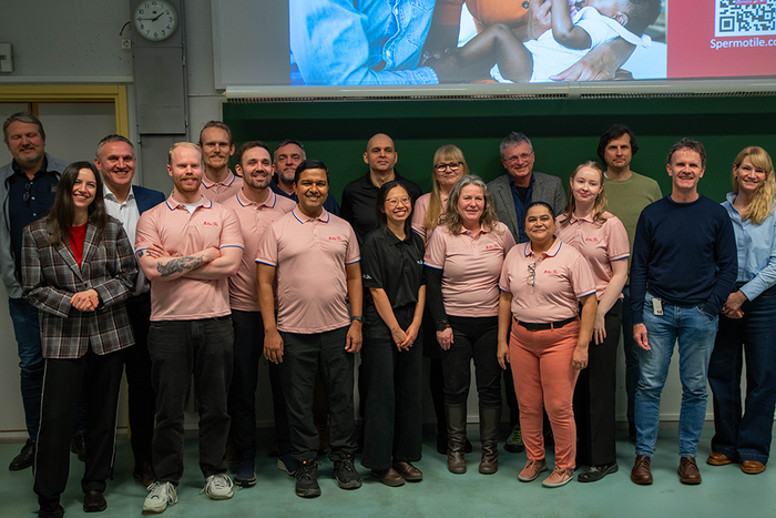
Spermotile: Advancing Male Fertility Through Multidisciplinary Collaboration Feb. 25, 2025 3:21 PM
-
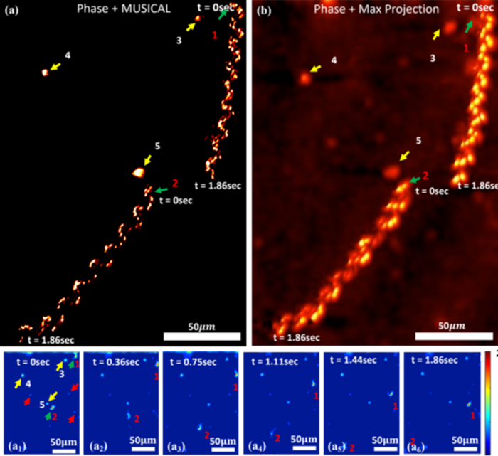
Unveiling the Mysteries of Sperm Motion Aug. 27, 2024 11:43 AM
-
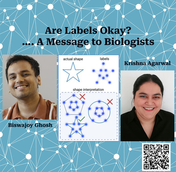
Are Labels Okay? - a message to biologists from DLN researchers June 14, 2023 2:00 PM
-
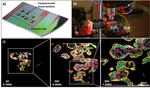
Photonic chip-based microscopy enables high throughput super-resolution microscopy June 13, 2022 9:26 PM
-
Super-resolution with lattice illumination Oct. 30, 2021 10:23 AM