index.html
SOLIS
Development of Real Volumetric Microscopy through Single Objective Light-Sheet Imaging System (SOLIS)
Highlights 2024
We published two papers in 2024 and have started with the final phase of the project, which will see engineered heart tissue imaging on our now fully functional oblique plane microscope.
Project overview
Project lead: Florian Ströhl
Institution: UiT - The Arctic University of Norway
Funding: RCN, 8M NOK
Duration: 01.07.2021 - 30.06.2025
Publications
-
Sommernes, Jon-Richard; Thiyagarajan, Dhivya Borra & Ströhl, Florian (2025). Rapid and gentle volumetric imaging of host–pathogen interactions in salmon skin cells using projective oblique plane microscopy. Scientific Reports. 15(1). doi: 10.1038/s41598-025-33371-2. Full text in Research Archive
-
Sommernes, Jon-Richard; Millett-Sikking, Alfred & Ströhl, Florian (2024). S-polarized light-sheets improve resolution and light-efficiency in oblique plane microscopy. Scientific Reports. 14(1). doi: 10.1038/s41598-024-53900-9. Full text in Research Archive Show summary
-
Davis, Samuel; Sommernes, Jon-Richard; hambura, sebastian; Riedel, Levin; Gil, Alejandro & Ikmi, Aissam [Show all 8 contributors for this article] (2024). Mesoscopic axially swept oblique plane microscope for the imaging of freely moving organisms with near-isotropic resolution. Biomedical Optics Express. 15(12), p. 6715–6724. doi: 10.1364/BOE.537262. Full text in Research Archive Show summary
-
Reischke, Marie; Vanderpoorten, Oliver & Ströhl, Florian (2023). Rapid prototyping of 1xN multifocus gratings via additive direct laser writing. Micro and Nano Engineering (MNE). 19. doi: 10.1016/j.mne.2023.100186. Full text in Research Archive Show summary
-
Ströhl, Florian (2023). Instant Volume Microscopy of Organoids with SOLIS. Microscopy Today. ISSN 1551-9295. 31(5), p. 23–26. doi: 10.1093/mictod/qaad064. Full text in Research Archive Show summary
-
Ströhl, Florian; Bruggeman, Ezra; Rowlands, Christopher; Wolfson, Deanna & Ahluwalia, Balpreet Singh (2022). Quantification of the NA dependent change of shape in the image formation of a z-polarised fluorescent molecule using vectorial diffraction simulations. Microscopy research and technique (Print). ISSN 1059-910X. 85(5), p. 2016–2022. doi: 10.1002/jemt.24060. Full text in Research Archive Show summary
-
Ströhl, Florian; Hansen, Daniel Henry; Grifo, Mireia Nager & Birgisdottir, Åsa Birna (2022). Multifocus microscopy with optically sectioned axial superresolution. arXiv.org. ISSN 2331-8422. doi: 10.48550/arXiv.2206.01257. Full text in Research Archive Show summary
-
Ströhl, Florian; Hansen, Daniel Henry; Grifo, Mireia Nager & Birgisdottir, Åsa birna (2022). Multifocus microscopy with optical sectioning and high axial resolution. Optica. 9(11), p. 1210–1218. doi: 10.1364/OPTICA.468583. Full text in Research Archive Show summary
-
Sommernes, Jon-Richard & Ströhl, Florian (2024). S-polarized light-sheets improve resolution and light efficiency in Oblique Plane Microscopy. Full text in Research Archive
-
Ströhl, Florian (2024). Brillouin Microscopy Development at the Arctic University of Norway. Full text in Research Archive
-
Ströhl, Florian (2024). The basics of optical alignment. Imaging and Microscopy. ISSN 2160-8881. Full text in Research Archive Show summary
-
Ströhl, Florian (2023). 3D+time microscopy of organoids: challenges and solutions. Full text in Research Archive
-
Ströhl, Florian (2023). Multifocus Microscopy with Optical Sectioning. Full text in Research Archive
-
Ströhl, Florian (2023). 3D+time microscopy of organoids: challenges and solutions. Full text in Research Archive
-
Sommernes, Jon-Richard & Ströhl, Florian (2022). 3D vectorial diffraction simulations of oblique plane microscopy systems with solid immersion tertiary objectives. Full text in Research Archive
-
Sommernes, Jon-Richard (2022). 3D vectorial diffraction simulations of oblique plane microscopy systems with solid immersion tertiary objectives. Full text in Research Archive
-
Tomas, Rolland & Ströhl, Florian (2022). Han bygger neste generasjons mikroskop. [Internet]. Forskning.no. Full text in Research Archive
-
Ströhl, Florian; Hansen, Daniel Henry; Sommernes, Jon-Richard; Reischke, Marie; Nàger, Mireia & Birgisdottir, Åsa birna (2022). Instant Volume Microscopy. Full text in Research Archive
-
Ströhl, Florian (2022). 3D&UV – exploring new concepts in advanced imaging. Full text in Research Archive
-
Ströhl, Florian (2022). 3D microscopy development at the Arctic University. Full text in Research Archive
-
Ströhl, Florian (2021). Optical transfer functions in inverted high-resolution light-sheet microscopy. Full text in Research Archive
About the project
SOLIS is a technology development project that will produce microscopy solutions for unprecedented capabilities in the field of high-speed volumetric fluorescence imaging with sub-micrometre resolution.
In this project, a completely new type of optical microscope will be developed that combines the best aspects of traditional confocal with light-sheet microscopy. Confocal microscopy is one of the most successful imaging methods in biomedicine and suitable to visualise the structure of cells and tissues in 3D space. However, as in confocal microscopy a single point is raster-sampled through the volume of interest, its acquisition speed is very slow. Furthermore, only a fraction of the fluorescent light generated inside the sample is actually captured by the microscope’s photodetector, which limits this technique’s application to living samples, which tolerate only a limited amount of light exposure.
Light-sheet microscopy, on the other hand, excels in imaging speed and low light-dose, both of which are critical for studies of living samples. In light-sheet microscopy, a thin slice of the sample is illuminated at a time and the produced fluorescence is imaged by a second lens, which is positioned at a right angle to the light-sheet. Although this arrangement is very efficient in collecting light from the sample, the requirement for a second lens limits access to the imaging volume and thus excludes the use of standard sample preparation protocols.
The new microscope, dubbed SOLIS (single-objective light-sheet imaging system), keeps the resolution and ease of use of confocal microscopy, i.e. only a single objective lens near the sample is required, while increasing its gentleness akin to light-sheet microscopy. Additionally, thanks to its unique optical design, SOLIS manages to surpass the imaging speed of classical light-sheet microscopy as, in essence, it records fully sectioned volumes in an instant.
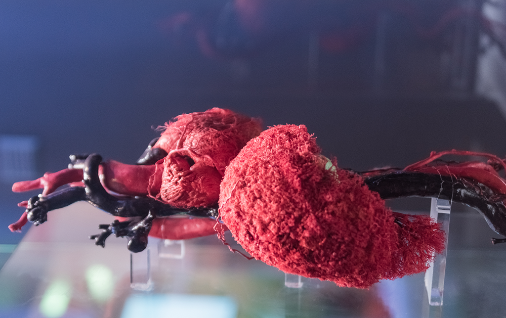
One application that will be enabled by the new system is the exploration of the repair mechanism of heart cells in 3D tissue models.